Cryo-Electron Microscopy
The Future of Cryo-Electron Microscopy
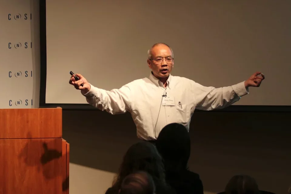
Hong Zhou, PhD
Director, Electron Imaging Center for NanoMachines (EICN)
Professor, Microbiology, Immunology & Molecular Genetics
Cryo-Electron Microscopy
Dr. Hong Zhou gains unprecedented insight into the mechanisms of infection by using cryo-electron microscopy to create some of the world’s most detailed 3-D models of viruses, bacteria, and other biological complexes. He achieved the first atomic resolution of a virus and rendered the clearest 3-D images of a herpes simplex virus type 1 ever recorded.
Dr. Zhou uses his models to design highly effective treatments that inhibit the precise structures and mechanisms pathogens use to infect hosts.
High-Resolution Models Lead to Better Infection Treatments
Dr. Zhou was training in atomic physics when a virus took his mother’s life and inspired him to study—and fight—infection. Driven to understand how infection works at the most detailed level possible, he turned to cryo-electron microscopy, a technique that uses beams of electrons to generate intricate images of biomolecules.
Dr. Zhou’s cryo-election models inform rational drug design—the development of treatments based on the structures and functions of specific viral targets.
Precision Antibiotics
Dr. Zhou and his team compared atomic resolution models of human ribosomes and parasite ribosomes, revealing a parasite-specific structure that antibiotics target. The finding gave researchers a new structural basis for developing novel, and potentially more effective, antibiotics. Read more in Structures and Stabilization of Kinetoplastid-Secific Split rRNAs Revealed by Comparing Leishmanial and Human Ribosomes, published in Nature.
Multi-Purpose Treatments
Dr. Zhou achieved the first atomic structural model of a herpesvirus capsid, the protein surrounding a virus’s nucleic acid. The virus he modeled is ubiquitous, maintains long-term latency, and possesses great genetic capacity, leading researchers to believe it could be used for gene delivery, oncolytic vectors, and vaccinations against other herpesviruses and even HIV/AIDS. Dr. Zhou’s models will guide the design of these therapeutic possibilities. Read more in Atomic Structure of the Human Cytomegalovirus Capsid with Its Securing Tegument Layer of Pp150, published in Science Magazine.
Can we use detailed viral models to develop better treatments?
Dr. Zhou believes we can push the limits of our measuring tools and techniques by combining cryo-electron microscopy, particle physics, and bioinformatics. He wants to learn more about how infection works and develop treatments based on solid structural evidence, trimming even more guesswork from medical practice.
"Why do I study herpes? We all have it; it is part of us, it has always been a part of us, and it will probably be part of us for years to come," says Dr. Zhou.
Dr. Zhou hopes cryo-electron microscopy leads to a future where we can study human cells in as much detail as we study virus cells. The detailed study of human cells will help us better understand and influence a range of biological processes, from cancer to infectious diseases.
“Instead of wondering if an antibiotic will work, we can look at the structure of the virus, see how the antibiotic interacts with it, and know for sure that it will work.”