• Gryphon Login
  • MyCourses
  • Alumni
  • UCLA Health
  • Contact Us
Cardiovascular

UCLA Cardiovascular Research Theme

Cardiovascular
  • About
    • Home
    • Overview
    • Theme Leadership
    • 2017 CV Theme Committee Members
    • Contact Information
    • Faculty Directory
    • What to Know About Cardiovascular Health

    Affiliated Programs

    • Medicine/cardiology
    • Adult Congenital Heart Disease
    • Physiology
    • Cardio-Thoracic Surgery
    • Anesthesiology
    • Pediatric Cardiology
    • UCLA Cardiovascular Symposium 2017
    • Leadership
    • Contact Us
    • Trainee Spotlight
    • Heart Heroes
    • Theme Committee Members
    • Cardiovascular Health
    • Newsletter
    • Seminar Series
  • Precision Health
  • Heart Failure & Regeneration
  • Arrhythmia & Sudden Death
  • Atherosclerosis & Vascular Biology
  • Seminars & Outreach
    • Seminar Announcements
  • Faculty Directory
  • Support Science
  • Gryphon Login
  • MyCourses
  • Alumni
  • UCLA Health
  • Contact Us

UCLA Cardiovascular Research Theme

Heart Failure & Regeneration

  1. Home
  2. Cardiovascular
  3. Heart Failure & Regeneration

Scarless Healing After Heart Attack

Share this
Cell fate mapping in the heart (Image courtesy of Ardehali Lab)

Cell fate mapping in the heart (Image courtesy of Ardehali Lab)

An advance that could significantly reduce the burden of chronic illness and death from cardiovascular disease would be to eliminate the scars that damage the heart muscle, impairing the heroic work of this organ.

Dr. Reza Ardehali, a UCLA associate professor of medicine (cardiology), heads a group of researchers exploring novel therapies to deliver stem cell-derived heart cells to replace scars formed after a heart attack.

Scarring: A Cause of Heart Failure

Scar tissue is a leading cause of arrhythmia, heart failure and sudden cardiac death.

In a major heart attack, a person loses an estimated 1 billion heart cells, which results in permanent scarring of the heart tissue. Loss of cell elasticity happens as part of aging as well, similar to how skin changes and sags over time.

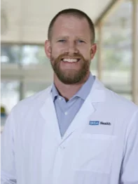
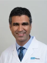
Dr. Reza Ardehali

Dr. Reza Ardehali

Scarring causes several functional problems for the heart:

  • When tissue dies, scars are how the body fills the gap. Initially, scarring is a beneficial part of the healing process.
  • Later, patches of collagen replace healthy cells that have a lot of elastic fibers. This process, known as fibrosis, makes tissue more rigid and with lung disease fibrosis makes it difficult to breathe.
  • A scar made up of dead heart cells cannot contribute to the contractile force of the heart – making it harder for the heart to beat.
  • Over time, the loss of elasticity has devastating consequences for heart function and the health of the patient.

“A scar in the heart takes the place of cardiomyocytes – heart cells that beat,” says Dr. Jake Lusis, professor of medicine, human genetics, microbiology, immunology and molecular genetics at UCLA. “At a minimum, this makes the heart less efficient. In the worst-case scenario, it can short-circuit the heart’s electrical system.”

Changing the Fate of a Scar

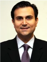
Dr. Arjun Deb

Dr. Arjun Deb

Reversing scarring, or preventing it in the first place, has been one of the major challenges of cardiovascular research, says Dr. Arjun Deb, an associate professor of medicine (cardiology) and senior author of a UCLA study published in 2014 in Nature.

The study found that the fibroblasts that form a scar also have the ability to regenerate healthy blood vessel cells in the heart. An overview of the study includes the following:

  • Investigators conducted experiments in mice in which scar-forming cells in the heart were genetically labeled.
  • The researchers discovered that many of the fibroblasts in the heart’s injured region changed into endothelial cells and contributed directly to blood vessel formation – a phenomenon they called mesenchymal to endothelial transition (MEndoT).
  • Investigators found that administering a small molecule to augment the process led to less scarring, allowing the heart to heal more completely.
  • Finding the switch that determines whether injured tissue will produce a scar or regenerate means that researchers can now attempt to manipulate that switch.
  • Investigators further discovered that once a heart attack occurs, there is a limited window in which therapy can coax scar-forming cells to produce blood cells instead.
  • This discovery suggests that after a heart attack, physicians can administer agents that will prevent scars from forming and promote cell regeneration.

Dr. Deb is currently looking for such an agent. “Critical time windows are important for other cardiac lifesaving drugs such as tPA (tissue plasminogen activator). We are working on identifying critical time windows for modulating scar tissue,” he says.

Calcification: A Long Term Consequence of Scarring

Aldons_Lusis.png

Jake Lusis, PhD 

In the process of seeking ways to regenerate, or “turn back the clock,” from heart damage, Dr. Deb and his group have studied the life cycle of scars. They have learned that the older scars get, the more they harden, or calcify.
The biology of scars and how they behave with time may be important for determining long-term heart function. Investigators have observed:

  • Scar-forming cells contribute to the calcification of scars.
  • These scars, in turn, can further contribute to cardiac dysfunction such as conduction system disturbances and heart blocks.
  • Critical pathways appear to regulate the ability of scar-forming cells to calcify scars. (Discovered in collaboration with the lab of Dr. Jake Lusis, vice chair of Human Genetics at UCLA.)

Dr. Deb says that there are remarkable similarities in the process of scarring in different organs after injury. His hope is that our findings and the therapeutic approaches we are developing can be used to treat scar tissue in other organs as well.

Generating New Cells for Ailing Hearts

Other UCLA researchers are working on ways to mend hearts through regeneration and cell therapy.

Dr. Ardehali treats patients with advanced heart disease, while also studying ways to cure or reverse heart disease. Currently, mechanical circulatory support devices and heart transplants are the last options for patients with end-stage heart disease. The limited therapies available to such patients motivated Dr. Ardehali to investigate a regenerative approach to restore heart function.

Stem Cells as Alternative Therapy

Dr. Ardehali believes it will one day be possible to offer alternative therapies by delivering stem cells to regenerate the damaged heart.
The process might work this way:

  • Clinicians would take a sample of skin cells from a patient.
  • The cells would be turned into pluripotent-like stem cells. This would basically endow these cells with the ability to become any tissue.
  • Cells would be engineered to become myocytes, or heart cells.
  • These laboratory-grown, individualized heart cells will be transplanted into a patient’s damaged heart at the site of damage.
  • The cells would repair the heart and regrow healthy tissue.

This technique could benefit patients with the following conditions:

  • Heart attack: Patients with heart attacks would be the first set of patients who would benefit from this advance.
  • Heart failure: The technique might help repair scars that are causing arrhythmias and contributing to heart failure.
  • Heart transplant: Ultimately, the technique would be a major therapy for patients with end-stage heart failure whose current options are limited to heart transplantation. In the future, instead of replacing the entire organ, cell transplantation can be used to repopulate the muscle that has been lost due to heart failure.

Dr. Ardehali and his team are actively working to make his vision a reality. His collaborators include UCLA stem cell biologists, cardiologists, bioengineers, developmental biologists and cardiac surgeons, among others.

Finding and Tracking Stem Cells in the Heart

Blood vessel network (Image courtesy of Iruela-Arispe Lab)

Blood vessel network (Image courtesy of Iruela-Arispe Lab)

Dr. Ardehali has identified a unique type of stem cells that can create heart cells and has successfully used new techniques to track transplanted cells via imaging. Recent discoveries include:

  • Cell markers: As detailed in the January 2016 issue of Stem Cell Reports, Dr. Ardehali has studied two specific markers that identify progenitor cells with the ability to generate both heart muscle and the blood vessels that support heart function. These cells:
    • May eventually help regenerate heart tissue
    • Successfully engraft when transplanted
    • Generate muscle tissue in the heart
  • Cell labeling and tracking: Another study published recently debuted a novel approach to imaging, labeling and tracking transplanted cells in the heart using MRI.
    • These techniques have allowed Dr. Ardehali and his colleagues to test cardiomyocyte transplantation in a pig, essentially a large animal model of human heart function.
    • UCLA is one of the few centers to have successfully carried out these studies in the pig model – an important step toward clinical implementation.

UCLA researchers have shown that although the rate of retention is very low – less than 10% of the cells engraft and integrate – some cells survive. Investigators have documented the cells’ differentiation into cardiac muscle and into vascular cells in the heart.

These therapies are still at an experimental level in the laboratory. Many hurdles still need to be overcome prior to clinical application. Still, Dr. Ardehali regards the progress to date as a “very promising first step to repopulate the heart in the hope of restoring its normal function.”

Like Us on Facebook Follow Us on Twitter Subscribe to Our Videos on YouTube Follow us on Instagram Connect with Us on LinkedIn Follow us on Pinterest Follow us on Flickr Follow us on Sharecare
Top 10 U.S. Medical Schools
  • Giving
  • Contact Us
  • Diversity
  • Emergency
  • Maps & Directions
  • Publications
  • Directory
  • Report Misconduct
  • Volunteer
  • Biomed Library
  • Newsroom
  • Smoke-Free
  • Sitemap
  • Terms of Use
  • Report Broken Links
Top 10 U.S. Medical Schools
Like Us on Facebook Follow Us on Twitter Subscribe to Our Videos on YouTube Follow us on Instagram Connect with Us on LinkedIn Follow us on Pinterest Follow us on Flickr Follow us on Sharecare