Stem Cells
Neural Stem Cells to Human Neurons
3D Structuring: brain balls
Rapid development in stem cell biology harbors enormous potential to provide insights into psychiatric disorders including autism. Here neural stem cells from patients can be coaxed into becoming neurons in a dish. But instead of settling for a flat, two-dimensional patch of cells, researchers are now able to create 3-D brain-like structures from the neural stem cells. Called brain balls, brains in a dish, or cerebral organoids - these clumps of cells are about the size of the head of a pin but reveal features of cell organization similar to the intact brain much more so than for cells in 2-D form. Brain balls can be used to model a disease process.
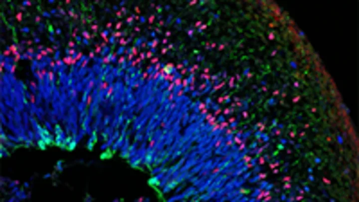
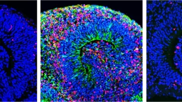
Cross section - Organoid
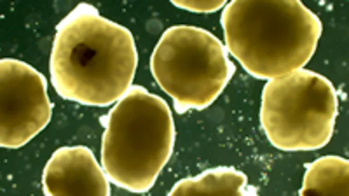
Organoids - in a dish
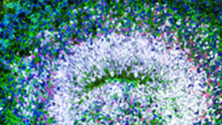
Cross section - Organoid
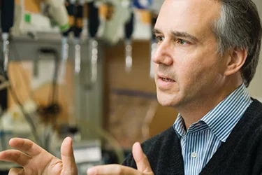
“What's exciting about this is that we can get a glimpse of what actually happens in early development.”
"We tend to talk about cells as individual entities. But in the nervous system, and in the development of neuropsychiatric disorders, cells have to communicate with one another," he says. "One thing we've not done a very good job with is coming up with ways to show the communication process."
Brain balls are made by taking neural stem cells and using chemicals to prompt the cells to form multi-layered tissue, activating the development programs that lead to the formation of different types of neurons and circuits. Researchers insert fluorescent markers into cells that are activated by light to follow what a cell is doing within the brain ball and to record the action with time-lapse imaging.
"What's exciting about this is that we can get a glimpse of what actually happens in early development," Dr. Novitch says. "And we can follow this out over time and actually see neurons forming. As the neurons form they start to form connections with one another. These structures are much closer to what you'd see in the real body" than cells growing in a dish in 2D. By comparison of the structure and function of neurons in these 3D brain like structures from patient cells to similar structures from unaffected individuals, researchers anticipate gaining insights into differences in cellular functions between them.
Disease in a dish
Below you see a montage of organoids, from left to right: before zika infection, after zika infection, and zika infection with a drug that we found blocks infection. The red here are dying cells.